
When researching NIPT, you often see terms like chromosome 1, chromosome 21, or “all chromosomes”—but why are they numbered in the first place? Many people ask themselves this exact question.
Human chromosomes are divided into autosomes (numbered 1 through 22) and sex chromosomes (X and Y chromosomes).
The autosomes are numbered 1 to 22 based on their physical size, arranged in descending order from the longest to the shortest.
A comprehensive “all-chromosome” test (chromosomes 1–22) screens the entirety of these autosomes, focusing on the regions that carry vital genetic information.
Because structural or numerical anomalies in these chromosomes can cause systemic health or developmental challenges, this screening plays a crucial role in evaluating the comprehensive health of the fetus.

What Is the Screening?
While conventional (standard) NIPT primarily targets trisomies 21, 18, and 13, Hiro Clinic’s full-chromosome aneuploidy screening represents an advanced, evolved diagnostic methodology.
Our approach comprehensively screens for numerical abnormalities (aneuploidies) across all chromosomes from 1 to 22, as well as the sex chromosomes, and provides detailed reports on any chromosome where an anomaly is detected. This genome-wide method has garnered significant clinical attention in recent years because it offers a substantially more comprehensive overview of genetic information.
When anomalies are detected on chromosomes other than 21, 18, and 13, the likelihood of fetal survival is often lower. Specifically, numerical aberrations involving autosomes outside of chromosomes 13, 18, and 21 can have profound, life-limiting consequences on fetal development. For this reason, comprehensive and exhaustive screening across the entire genome carries vital clinical significance.
International prenatal screening guidelines recognize the value of broader, more comprehensive chromosomal anomaly screening, which includes information regarding sex chromosomes that conventional testing methodologies often fail to capture. While standard prenatal screening has historically focused primarily on the three major autosomes, clinical data indicates that even when those specific autosomes are unaffected, anomalies can still arise within the sex chromosomes.
Statistical data shows that these expanded conditions yield positive diagnostic results in approximately 0.56% of chorionic villus samplings (CVS) (roughly 1 in 200 cases) and approximately 0.05% of amniocentesis procedures (roughly 1 in 2,000 cases). Because these anomalies could potentially be missed using conventional targeted methods, genome-wide full-chromosome aneuploidy screening has become an essential tool in significantly reducing diagnostic uncertainty.
The Difference Between Monosomy and Trisomy
When a typical pair of chromosomes gains an extra chromosome, resulting in a total of three, it is referred to as a trisomy. Conversely, when a pair loses a chromosome, leaving only a single one, it is called a monosomy.
Why is Age 35 Set as the Standard for Pregnant Individuals?
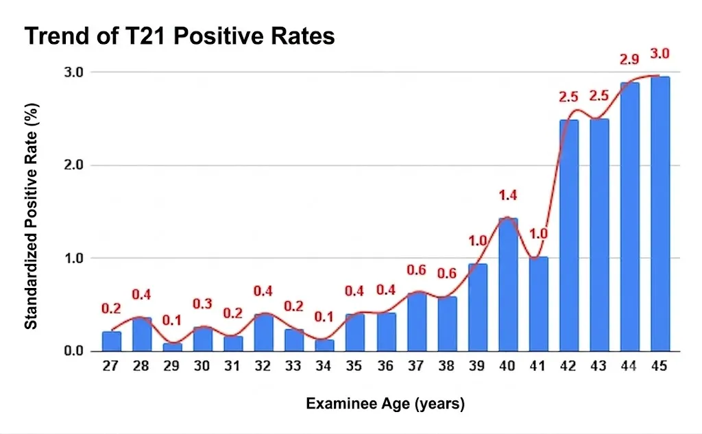
Historically, standard NIPT (Non-Invasive Prenatal Testing) was only available to pregnant individuals aged 35 and older. The reason for this threshold is that the incidence rate of autosomal numerical abnormalities shifts significantly around the age of 35.
At Hiro Clinic, we have compiled and analyzed screening outcomes from patients ranging in age from 19 to 51 who underwent NIPT. Advanced maternal age is clinically defined as 35 years or older.
Out of 809 patients who received a positive screening result for Trisomy 21 (Down syndrome), 613 were aged 35 or older. As demonstrated in the data, the frequency of positive results increases progressively with maternal age.
Positive Rates of Full-Chromosomal Aneuploidy in Chorionic Villus Sampling (CVS) and Amniocentesis
| Trisomies 21, 18, and 13 | Other Trisomies | |
|---|---|---|
| Number of Samples in Chorionic Villus Sampling (CVS) |
5.06% (532 out of 10,511 cases) |
0.56% (59 out of 10,511 cases) |
| Number of Samples in Amniocentesis |
1.02% (748 out of 73,268 cases) |
0.05% (30 out of 73,268 cases) |
| Number of Samples in Both CVS and Amniocentesis Procedures |
2.1% (235 out of 11,066 cases) |
0.12% (13 out of 11,066 cases) |
1. Konialis C, Pangalos C. Dilemmas in prenatal chromosomal diagnosis revealed through a single center’s 30 years’ experience and 90,000 cases. Fetal Diagn Ther 2015; 38(3):218-232.
2. Comas C, Echevarria M, Rodríguez I, Serra B, Cirigliano V. Prenatal invasive testing: a 13-year single institution experience. J Matern Fetal Neonatal Med 2014 Aug;27(12):1209-12.
Reported Positive Cases by Trisomy Type in Full-Chromosomal Aneuploidy Screening(Excluding Mosaicism)
| Trisomy 1 | Blighted ovum |
|---|---|
| Trisomy 2 | Non-viable |
| Trisomy 3 | No positive cases reported |
| Trisomy 4 | Rare: Non-viable |
| Trisomy 5 | Non-viable |
| Trisomy 6 | Non-viable |
| Trisomy 7 | Non-viable |
| Trisomy 8 | Non-viable |
| Trisomy 9 | Live births documented |
| Trisomy 10 | Non-viable |
| Trisomy 11 | Non-viable |
| Trisomy 12 | Non-viable |
|---|---|
| Trisomy 13 | Live births documented |
| Trisomy 14 | Non-viable |
| Trisomy 15 | Live births documented |
| Trisomy 16 | Non-viable |
| Trisomy 17 | Non-viable |
| Trisomy 18 | Live births documented |
| Trisomy 19 | Extremely rare: Non-viable |
| Trisomy 20 | Live births documented |
| Trisomy 21 | Live births documented |
| Trisomy 22 | Live births documented |
Frequently Asked Questions About Full-Chromosomal Screening
Q: Does full-chromosomal screening detect every type of fetal chromosomal abnormality?
A: Full-chromosomal screening specifically checks for “numerical abnormalities” (aneuploidies), such as trisomies (where there are three copies of a chromosome instead of a pair) or monosomies (where there is only a single copy) across autosomes 1 to 22 and the sex chromosomes.
To evaluate structural abnormalities—such as deletions or duplications of chromosomal segments—rather than just numerical shifts, an expanded screening for structural copy number variants (partial deletions and duplications)* is required.
For more details regarding structural variations, please refer to the section titled “What Are Partial Chromosomal Deletion and Duplication Disorders?“
*Please note: This structural analysis is limited to chromosomal aberrations spanning 5 megabases (Mb) or greater.