228 recessive genetic disorders identified by Hiro Clinic

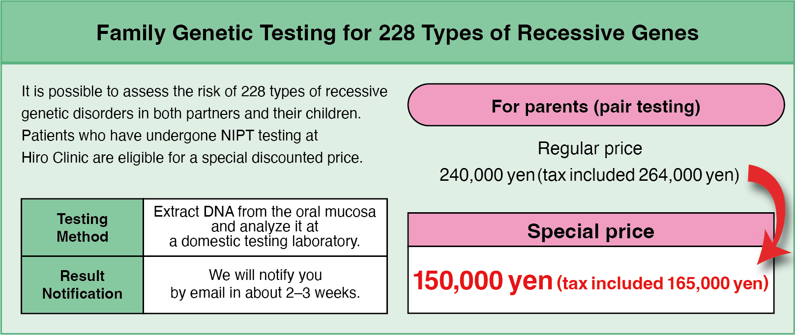
Regarding the Test for 228 Types of Recessive Genetic Disorders

At Hiro Clinic, we offer a test that can examine 228 genes associated with severe recessive genetic disorders before birth.

Recessive genetic disorders are conditions that occur when there is an abnormality in the genes.
In this test, to determine whether both parents carry gene abnormalities, genes are extracted and analyzed from the oral mucosa of the mother and father.
By testing whether both parents have abnormalities in the same gene location, it is possible to predict the risk of the fetus having a severe genetic disorder.
For example, if both parents have abnormalities in the same gene, the child is at higher risk of developing a recessive genetic disorder (about 25%). If the test result is positive, amniocentesis is necessary as a confirmatory test.

So, what kind of test is conducted for such individuals? Also, what kind of disorders are identified?

World Class Next Generation NIPT

New users here

新規登録

Registered users here

マイーページ

24hours reservation available

夫婦の写真PC
夫婦の写真MOBILE

It is recommended that couples/couples consider genetic testing

Hiro Clinic recommends that you undergo 228 different recessive gene tests. This allows you to find out about conditions that may be inherited by your child before birth. Knowing this information can be useful for future health and family planning, enabling you to plan early measures and support.

Benefits

Comprehensive risk assessment
The genetic information of the couple or couples can be used to extensively assess and take necessary action for diseases that may be inherited by their children.

Family planning support
Support decision-making to take genetic risk into account in future family planning.

Optimising preventive measures
Optimising preventive measures: individualised preventive measures and early intervention can be planned based on identified genetic risks.

赤ちゃんの写真PC
赤ちゃんの写真MOBILE

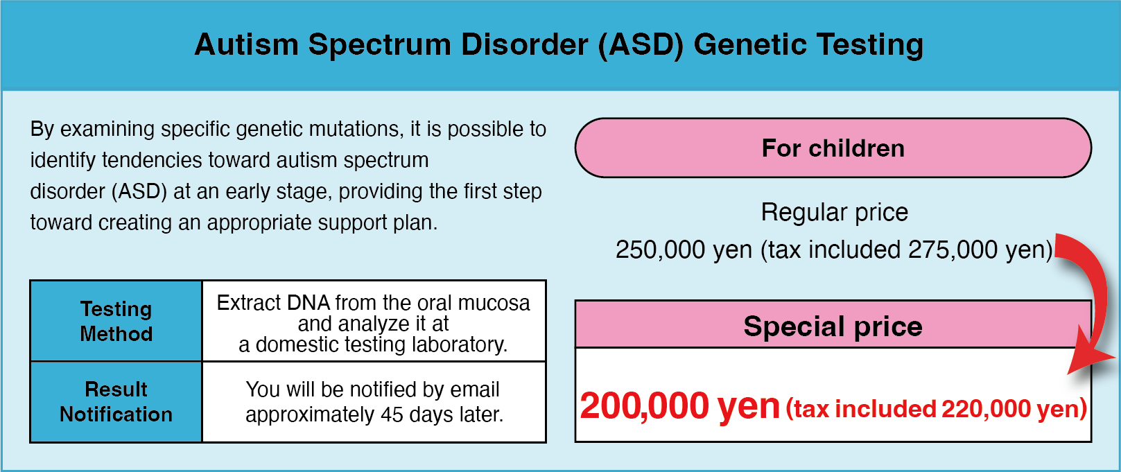
We recommend that you consider taking a genetic test for your newborn baby

Having your newborn baby undergo recessive genetic testing is one way to support your child’s health and future well-being. The test allows for early identification of genetic risks and, if necessary, appropriate preventive measures and treatment can be initiated immediately. At Hiro Clinic, we prioritise your child’s safety and comfort and use state-of-the-art technology to offer a painless ‘just rub the stick of the kit on the buccal mucosa (cheek in the mouth)’ test.

Benefits

Early detection
Screening for 228 recessive genetic conditions to detect potential risks to your child’s health at an early stage.

Individualised health care
Based on the test results, specific health care and treatment needs can be planned for your child.

Family security
Clear information about your child’s health status can help ease family anxiety.

Special limited price for those who have undergone Hiro Clinic NIPT
Hiro Clinic offers 228 recessive genetic tests at a special price to those who have undergone NIPT.
More information

If you have not undergone NIPT, don’t worry
228 recessive genetic tests are also available for those who have not undergone NIPT at Hiro Clinic.
More information

Inspection process

Step 1

Apply for an inspection at the clinic

The consent form of the pregnant woman and her partner will be included in the application
The test kit is shipped to your home (takes about one week from the visit)
※Pregnant women and their partners must come to the clinic together
Step 2

The test kit is delivered to your home

Test kits are enclosed with test sets for pregnant women and their partners
Step 3

Do the test at home

Rub the shaft part of the test kit on the buccal mucosa (oral cheek) to collect cells
Step 4

Return of test kits

Place the collected specimen in a letter-pack and post it in your local post box
Step 5

Inspection report

We will report back to you by email in about a month
※This is a separate report from the national tests conducted by drawing blood from pregnant women
※Tests are reported approximately one month after the returned test kit arrives at the laboratory

Optional Fees for 228 Types of Recessive Gene Tests
Available

228 Types of Recessive Gene Tests asd Types of Recessive Gene Tests

How recessive genetic disorders develop

Humans have two chromosomes, one of maternal origin and one of paternal origin.

A recessive genetic disorder is a disease that develops when two chromosomes are abnormal in the same place. If one of the chromosomes of maternal or paternal origin is abnormal, but the other chromosome is not abnormal, the disease does not occur. In that case, the individual is said to be a carrier of the relevant genetic disorder.

Carriers are the ones who have been told since ancient times that they should not have children by blood.

For example, if there is a rare disease that affects only one in 40 000 people, it is estimated that there is one carrier in every 100 people.

This is because the calculation is ‘1 in 100 x 1 in 100 x 1 in 4 = 1 in 40,000’.

If we test 100 places for one disease in 40,000 people, this means that theoretically everyone could be a carrier of one disease.

So what happens if our test shows an abnormality in the same place in both parents’ genes?

Answer is

  • One in four foetuses will develop the disease.
  • One in two people will be a carrier.
  • One in four individuals will be normal.

How can it be found out?

At present, the most accurate one is the amniotic fluid test.

This amniotic fluid test requires genetic analysis. Tokyo Hygienic Laboratory carries out this analysis in collaboration with Medicover in Cyprus.

If you have amniotic fluid testing support, the test usually costs less than 200 000 yen, so there is no cost burden.
Please contact Hiro Clinic for more information on amniotic fluid testing.

The 228 recessive genetic disorders are the following diseases.

Hiro Clinic NIPT can tell you about
Recessive genetic disorders

Disease Name Genes Tested at Our Hospital Chromosomes that arise Detailed Information
3-Hydroxy-3-Methylglutaryl-Coenzyme A Lyase Deficiency HMGCL 1p36.1 More Information
Leydig Cell Hypoplasia [Luteinizing Hormone Resistance] LHCGR 2p16.3 More Information
3-Methylcrotonyl-CoA Carboxylase Deficiency 1 MCCC1 3q27.1 More Information
Limb Girdle Muscular Dystrophy, Type 2E SGCB 4q12 More Information
3-Methylcrotonyl-CoA Carboxylase Deficiency 2 MCCC2 5q13.2 More Information
Lipoamide Dehydrogenase Deficiency [Maple Syrup Urine Disease, Type 3] DLD 7q31.1 More Information
Abetalipoproteinemia MTTP 4q23 More Information
Lipoprotein Lipase Deficiency LPL 8p21.3 More Information
Acyl-CoA Oxidase I Deficiency ACOX1 17q25.1 More Information
Omenn Syndrome (RAG2-related) RAG2 11p12 More Information
Chorea-acanthocytosis VPS13A 9q21.2 More Information
Ornithine Aminotransferase Deficiency OAT 10q26.13 More Information
Choroideremia, X-Linked CHM Xq21.2 More Information
Ornithine Translocase Deficiency Hyperornithinemia-Hyperammonemia -Homocitrullinuria (HHH) Syndrome] SLC25A15 13q14.11 More Information
Citrin Deficiency SLC25A13 7q21.3 More Information
Pendred Syndrome SLC26A4 7q22.3 More Information
Combined Oxidative Phosphorylation Deficiency 3 TSFM 12q14.1 More Information
Peroxisome Biogenesis Disorders Zellweger Syndrome Spectrum (PEX1-related) PEX1 More Information
Congenital Disorder of Glycosylation, Type 1A (PMM2-related) PMM2 16p13.2 More Information
Peroxisome Biogenesis Disorders Zellweger Syndrome Spectrum (PEX2-related) PEX2 More Information
Hereditary Fructose Intolerance ALDOB 9q31.1 More Information
Peroxisome Biogenesis Disorders Zellweger Syndrome Spectrum (PEX1-related) PEX1 7q21.2 More Information
Homocystinuria, Type cblE MTRR 5p15.31 More Information
Severe Combined Immunodeficiency, X-Linked IL2RG Xq13.1 More Information
Hydrolethalus Syndrome HYLS1 11q24.2 More Information
Sickle-Cell Disease HBB 11p15.4 More Information
Inclusion Body Myopathy, Type 2 GNE 9p13.3 More Information
Sjögren-Larsson Syndrome ALDH3A2 17p11.2 More Information
Isovaleric Acidemia IVD 15q15.1 More Information
Steroid-Resistant Nephrotic Syndrome NPHS2 1q25.2 More Information
Methylmalonic Aciduria, Type Mut(0) MMACHC 1p34.1 More Information
Stuve-Wiedemann Syndrome LIFR 5p13.1 More Information
Methylmalonic Aciduria and Homocystinuria, Type cblD MMADHC 2q23.2 More Information
Bardet Biedl Syndrome (BBS12-related) BBS12 4q27 More Information
Mucopolysaccharidosis, Type II [Hunter Syndrome], X-Linked IDS Xq28 More Information
Beta Thalassemia HBB 11p15.4 More Information
Mucopolysaccharidosis, Type IIIC [Sanfilippo C] HGSNAT 8p11.21-p11.1 More Information
Biotinidase Deficiency BTD 3p25.1 More Information
Multiple Sulfatase Deficiency SUMF1 3p26 More Information
Canavan Disease ASPA 17p13.2 More Information
Primary Ciliary Dyskinesia (DNAH5-related) DNAH5 5p15.2 More Information
Carpenter Syndrome RAB23 6p12.1-p11.2 More Information
Primary Ciliary Dyskinesia (DNAI1-related) DNAI1 14q24.3 More Information
Glycogen Storage Disease, Type 1A(BBS12-related) G6PC 17q21 More Information
Primary Hyperoxaluria, Type 3 HOGA1 10q24.2 More Information
Glycogen Storage Disease, Type 1B SLC37A4 11q23.3 More Information
Pycnody sostosis CTSK 1q21.3 More Information
Glycogen Storage Disease, Type 3 AGL 1p21.2 More Information
Pyruvate Dehydrogenase Deficiency (PDHB-Related) PDHB 3p14.3 More Information
Glycogen Storage Disease, Type 7 BCS1L 2q35 More Information
Aicardi-Goutières Syndrome SAMHD1 20q11.23 More Information
GRACILE Syndrome BCS1L 2q35 More Information
Alport Syndrome, X-Linked COL4A5 Xq22.3 More Information
Long Chain 3-Hydroxyacyl-CoA Dehydrogenase Deficiency HADHA 2p23 More Information
Alstrom Syndrome ALMS1 2p13.1 More Information
Lysinuric Protein Intolerance SLC7A7 14q11.2 More Information
Andermann Syndrome SLC12A6 15q14 More Information
Maple Syrup Urine Disease, Type 1B BCKDHB 6q14.1 More Information
Aromatase Deficiency CYP19A1 15q21.2 More Information
Methylmalonic Acidemia (MMAA-related) MMAA 4q31.21 More Information
Congenital Neutropenia (HAX1-related) HAX1 1q21.3 More Information
Methylmalonic Aciduria and Homocystinuria, Type cblC MMAA 4q31.21
Crigler Najjar Syndrome, Type I UGT1A1 2q37.1 More Information
Peroxisome Biogenesis Disorders Zellweger Syndrome Spectrum (PEX2-related) PEX2 8q21.13 More Information
Cystic Fibrosis CFTR 7q31.2 More Information
Phenylketonurea PAH 12q23.2 More Information
Factor XI Deficiency F11 4q35.2 More Information
Pontocerebellar Hypoplasia, Type 1A VRK1 14q32.2 More Information
Familial Dysautonomia IKBKAP 9q31.3 More Information
Pontocerebellar Hypoplasia, Type 2D SEPSECS 4p15.2 More Information
Joubert Syndrome, Type 2 LAMC2 1q25.3 More Information
Pontocerebellar Hypoplasia, Type 2E VPS53 17p13.3 More Information
Junctional Epidermolysis Bullosa, Herlitz Type LAMC2 1q25.3 More Information
Tay-Sachs Disease HEXA 15q23 More Information
Lamellar Ichthyosis, Type 1 TGM1 14q12 More Information
Usher Syndrome, Type 1F PCDH15 10q21.1 More Information
Leber Congenital Amaurosis (LCA5-related) LCA5 6q14.1 More Information
Usher Syndrome, Type 3 CLRN1 3q25.1 More Information
Leigh Syndrome, French-Canadian Type LRPPRC 2p21 More Information
Wolman Disease LIPA 10q23.31 More Information
Myotubular Myopathy, X-Linked MTM1 Xq28 More Information
3-Methylglutaconic Aciduria, Type 3 [Costeff Syndrome], OPA3 More Information
Navajo Neurohepatopathy [MPV17-related Hepatocerebral Mitochondrial DNA Depletion Syndrome] MPV17 2p23.3 More Information
Abetalipoproteinemia MTTP More Information
Acute Infantile Liver Failure (TRMU-related) TEMU More Information
Neuronal Ceroid Lipofuscinosis (CLN8-related) CLN8 8p23 More Information
Argininosuccinate Lyase Deficiency, ASL ASL More Information
Neuronal Ceroid Lipofuscinosis (MFSD8-related) MFSD8 4q28.2 More Information
Ataxia-Telangiectasia, ATM ATM More Information
Neuronal Ceroid Lipofuscinosis (TPP1-related) TPP1 11p15.4 More Information
Autosomal Recessive Spastic Ataxia of Charlevoix- Saguenay, SACS SACS
Nijmegen Breakage Syndrome NBN 8q21.3 More Information
Bare Lymphocyte Syndrome (CIITA-related), CIITA CIITA More Information
Retinal Dystrophy (RLBP1-related) [Bothnia Retinal Dystrophy] RLBP1 15q26.1 More Information
“Combined Oxidative Phosphorylation Deficiency 1, GFM1” GFM1 More Information
Congenital Finnish Nephrosis, NPHS1 NPHS1 More Information
Retinitis Pigmentosa 25 (EYS-related) EYS 6q12 More Information
“Congenital Myasthenic Syndrome (RAPSN-related), RAPSN” RAPSN More Information
Retinitis Pigmentosa 59 (DHDDS-related) DHDDS 1p36.11 More Information
Corneal Dystrophy and Perceptive Deafness, SLC4A11 SLC4A11 More Information
Sanfilippo Syndrome, Type D [Mucopolysaccharidosis IIID] GNS 12q14.3 More Information
Creatine Transporter Defect [Cerebral Creatine Deficiency Syndrome 1] X-Linked, SLC6A8 SLC6A8 More Information
Severe Combined Immunodeficiency, Type Athabaskan DCLRE1C 10p13 More Information
Cystinosis, CTNS CTNS More Information
Arthrogryposis Mental Retardation Seizures SLC35A3 1p21.2 More Information
Duchenne Muscular Dystrophy, X-linked, DMD l DMD More Information
Arthrogryposis Mental Retardation Seizures SLC35A3 1p21.2 More Information
Emery-Dreifuss Muscular Dystrophy 1, X-Linked, EMD EMD More Information
Asparagine Synthetase Deficiency ASNS 7q21.3 More Information
Fabry Disease, X-Linked, GLA GLA More Information
Aspartylglycosaminuria AGA 4q34.3 More Information
Familial Mediterranean Fever, MEFV MEFV More Information
Autosomal Recessive Polycystic Kidney Disease PKHD1 6p12.3-p12.2 More Information
Galactosemia, GALT u GALT More Information
Bardet-Biedl Syndrome (BBS1-related) BBS1 11q13.2 More Information
Hemochromatosis, Type 3 (TFR2-related), TFR2 TFR2 More Information
Fanconi Anemia, Type C FANCC 9q22.32 More Information
Hermansky-Pudlak Syndrome (HPS3-related), HPS3 HPS3 More Information
Fanconi Anemia, Type G FANCG 9p13.3 More Information
Hypophosphatasia (ALPL-related), ALPL ALPL More Information
“3-Phosphoglycerate Dehydrogenase Deficiency, PHGDH” PHGDH
Gaucher Disease GBA 1q21 More Information
Glutaric Acidemia, Type 2A ETFA 15q24.2-q24.3 More Information
Glycine Encephalopathy (GLDC-related) GLDC 9p24.1 More Information
Leukoencephalopathy with Vanishing White Matter EIF2B5 3q27.1 More Information
Disease Name Genes Tested at Our Hospital Detailed Information
Autoimmune Polyglandular Syndrome, Type 1, AIRE AIRE More Information
Peroxisome Biogenesis Disorders Zellweger Syndrome Spectrum (PEX1-related), PEX1 PEX1
Bartter Syndrome (BSND-related), BSND BSND
Pituitary Hormone Deficiency, Combined 3, LHX3 LHX3
Cartilage-Hair Hypoplasia, RMRP RMRP More Information
Primary Ciliary Dyskinesia, DNAI1-related, DNAI1 DNAI1 More Information
Citrullinemia, Type 1, ASS1 ASS1
Primary Hyperoxaluria, Type 2, GRHPR GRHPR
Congenital Disorder of Glycosylation, Type 1B, MPI MPI More Information
“Pyruvate Dehydrogenase Deficiency (PDHB-related), PDHB” PDHB
“Congenital Insensitivity to Pain with Anhidrosis, NTRK1” NTRK1 More Information
Retinal Dystrophy (RLBP1-related) [Bothnia Retinal Dystrophy], RLBP RLBP
Corticosterone Methyloxidase Deficiency, CYP11B2 CYP11B2 More Information
Retinitis Pigmentosa 26, CERKL CERKL More Information
D-Bifunctional Protein Deficiency, HSD17B4 HSD17B4 More Information
Rhizomelic Chondrodysplasia Punctata, Type 1, PEX7 PEX7
“Dystrophic Epidermolysis Bullosa (COL7A1-related), COL7A1” COL7A1 More Information
Salla Disease, SLC17A5 SLC17A5
Segawa Syndrome, (TH-related), TH TH
Enhanced S-Cone Syndrome, NR2E3 NR2E3 More Information
Sickle-Cell Disease, HBB l HBB l
Factor IX Deficiency, X-Linked, F9 F9
Tay-Sachs Disease, HEXA u HEXA遺伝子
Familial Nephrogenic Diabetes Insipidus (AQP2- related), AQP2 AQP2 More Information
Wilson Disease, ATP7B ATP7B
Glycogen Storage Disease, Type 4, GBE1 GBE1 More Information
Junctional Epidermolysis Bullosa, Herlitz type, LAMC2 LAMC2
6-Pyruvoyl-Tetrahydropterin Synthase (PTPS) Deficiency, PTS PTS More Information
Lamellar Ichthyosis, Type 1, TGM1 TGM1
Achromatopsia (CNGB3-related), CNGB3 CNGB3
Leigh Syndrome, French-Canadian Type, LRPPRC LRPPRC
Adrenoleukodystrophy, X-Linked, ABCD1 ABCD1 More Information
Limb-Girdle Muscular Dystrophy, Type 2A, CAPN3 CAPN3
Alport Syndrome (COL4A3-related), COL4A3 COL4A3 More Information
Limb-Girdle Muscular Dystrophy, Type 2D, SGCA SGCA
Ataxia with Vitamin E Deficiency, TTPA TTPA More Information
Lipoid Adrenal Hyperplasia, STAR STAR
Batten Disease (CLN3-related), CLN3 CLN3
“Medium Chain Acyl-CoA Dehydrogenase Deficiency, ACADM u” ACADM u
Bloom Syndrome, BLM u BLM More Information
Metachromatic Leukodystrophy (PSAP-related) PSAP PSAP
Carnitine Palmitoyltransferase II Deficiency, CPT2 CPT2 More Information
Methylmalonic Aciduria and Homocystinuria, Type cblC, MMACHC MMACHC
Cerebrotendinous Xanthomatosis, CYP27A1 CYP27A1 More Information
Microphthalmia/Anophthalmia (VSX2-related), VSX2 VSX2 More Information
Chronic Granulomatous Disease, X-Linked, CYBB CYBB More Information
Mitochondrial Complex 1 Deficiency (NDUFS6-related), NDUFS6 NDUFS6
Combined Malonic and Methylmalonic Aciduria, ACSF3 More Information
Mucolipidosis III Gamma, GNPTG GNPTG
“Mucopolysaccharidosis, Type IIIB [Sanfilippo B], NAGLU” NAGLU
Combined Pituitary Hormone Deficiency 2, PROP1 PROP1 More Information
Mucopolysaccharidosis, Type IX, HYAL1 HYAL1
Congenital Disorder of Glycosylation Type 1C, ALG6 ALG6 More Information
“Congenital Myasthenic Syndrome (CHRNE-related), CHRNE” CHRNE More Information
Neuronal Ceroid Lipofuscinosis (CLN6-related), CLN6 CLN6
“Congenital Myasthenic Syndrome (CHRNE-related), CHRNE” CHRNE More Information
Niemann-Pick Disease, Types A/B, SMPD1 u SMPD1
Congenital Neutropenia (VPS45-related), VPS45 VPS45 More Information
Pontocerebellar Hypoplasia, RARS2-related, RARS2 RARS2
CRB1-related Retinal Dystrophies, CRB1 CRB1 More Information
Primary Ciliary Dyskinesia, DNAI2-related, DNAI2 DNAI2
Deafness, Autosomal Recessive 77, LOXHD1 LOXHD1 More Information
Pyruvate Dehydrogenase Deficiency, X-Linked, PDHA1 PDHA1
Ehlers-Danlos Syndrome, Type VIIC, ADAMTS ADAMTS More Information
Retinitis Pigmentosa 28, FAM161A FAM161A
Ethylmalonic Encephalopathy, ETHE1 ETHE1 More Information
Rhizomelic Chondrodysplasia Punctata, Type 3, AGPS AGPS
Factor V Leiden Thrombophilia, F5 F5
Sandhoff Disease, HEXB HEXB
Familial Hypercholesterolemia (LDLR-related), LDLR LDLR
Tyrosinemia, Type 1, FAH FAH
“Galactokinase Deficiency [Galactosemia, Type II], GALK1” GALK1
Usher Syndrome, Type 2A, USH2A USH2A
Glutaric Acidemia, Type 1, GCDH GCDH
Juvenile Retinoschisis, X-Linked, RS1 RS1
Glycine Encephalopathy (AMT-related), AMT AMT
Limb-Girdle Muscular Dystrophy, Type 2B, DYSF DYSF
“Glycogen Storage Disease, Type 2 [Pompe Disease], GAA” GAA
Megalencephalic Leukoencephalopathy with Subcortical Cysts, MLC1 MLC1
“Glycogen Storage Disease, Type 5 [McArdle Disease], PYGM” PYGM
Mitochondrial Complex 1 Deficiency (ACAD9-related), ACAD9 ACAD9
Hemochromatosis, Type 2A, HFE2 HFE2
Mitochondrial Myopathy and Sideroblastic Anemia (MLASA1), PUS1 PUS1
Hermansky-Pudlak Syndrome (HPS1-related), HPS1 HPS1
Mucolipidosis, Type IV, MCOLN1 u MCOLN1 u
Homocystinuria (CBS-related), CBS CBS
N-acetylglutamate Synthase Deficiency, NAGS NAGS
Hypohidrotic Ectodermal Dysplasia, X-Linked, EDA EDA
Niemann-Pick Disease, Type C1/D, NPC1 NPC1
Krabbe Disease, GALC GALC
Non-Syndromic Hearing Loss (GJB2-related, GJB6-related), GJB2, GJB6 GJB2、GJB6 More Information
Leber Congenital Amaurosis, Type CEP290, CEP290 CEP290
Ornithine Aminotransferase Deficiency, OAT OAT
Limb-Girdle Muscular Dystrophy, Type 2C, SGCG SGCG
Phenylketonurea, PAH u PAH u
Lipoamide Dehydrogenase Deficiency [Maple Syrup Urine Disease, Type 3], DLD DLD
Primary Hyperoxaluria, Type 1, AGXT AGXT
Long Chain 3-Hydroxyacyl-CoA Dehydrogenase Deficiency, HADHA HADHA
Renal Tubular Acidosis and Deafness (ATP6V1B1- related), ATP6V1B ATP6V1B
Meckel-Gruber Syndrome, Type 1, MKS1 MKS1
Retinitis Pigmentosa, X-linked, RPGR RPGR
Metachromatic Leukodystrophy (ARSA-related), ARSA ARSA
Roberts Syndrome, ESCO2 ESCO2
Methylmalonic Aciduria (MMAB-related), MMAB MMAB
Schimke Immunoosseous Dysplasia, SMARCAL1 SMARCAL1
Methylmalonic Aciduria, Type mut(0), MMUT MMUT
Smith-Lemli-Opitz Syndrome, DHCR7 u DHCR7 u
Mitochondrial Complex 1 Deficiency (NDUFAF5- related), NDUFAF5 NDUFAF5
Stuve-Wiedemann Syndrome, LIFR HSPG2
Mucolipidosis II/III, GNPTAB GNPTAB
Usher Syndrome, Type 1C, USH1C USH1C
“Myoneurogastrointestinal Encephalopathy (MNGIE), TYMP” TYMP
Zellweger Spectrum Disorders, (PEX6-related), PEX6 PEX6
Navajo Neurohepatopathy [MPV17-related Hepatocerebral Mitochondrial DNA Depletion Syndrome], MPV17 MPV17
Zellweger Spectrum Disorders (PEX10-related), PEX10 PEX10
Neuronal Ceroid Lipofuscinosis (CLN5-related), CLN5 CLN5
Neuronal Ceroid Lipofuscinosis (PPT1-related), PPT1 PPT1
Niemann-Pick Disease, Type C2, NPC2 NPC2
“Odonto-Onycho-Dermal Dysplasia / Schopf-Schulz- Passarge Syndrome, WNT10A” WNT10A
Ornithine Transcarbamylase Deficiency, OTC OTC

What are recessive genetic disorders at Hiro Clinic?

Hiro Clinic can detect 228 serious recessive genetic disease gene associations before birth.

This involves extracting and analysing genes from the buccal mucosa of the mother and father and combining them to detect serious genetic disorders in the foetus.

Serious genetic disorders are very rare, but even for a recessive genetic disorder that affects 1 in 40 000 people, the number of carriers of the disease (individuals with an abnormality in one of the two chromosomes but without the disease) is 1 in 100.

The HIRO Clinic considers the test to be useful in identifying carrier genes and suspecting a link between 228 different recessive genetic disorders based on their combination.

The unique feature of this test is that it detects all the combinations of genes that have been previously considered to be genetic abnormalities and determines whether the genetic changes are indeed pathogenic.

Genes differ from individual to individual. This is often the case for important genes, and their diversity makes each person different. However, they can also be pathogenic in their variation.

Gene combinations are shared in databases around the world, which are searched to see if any of the 228 gene variants are pathogenic.

When the mother and father are pathogenic at the same site, there is a high frequency risk of the child developing the disease. However, it is not enough just to look at the genetic change. It is necessary to determine (annotate) whether the genetic change is really abnormal. This is a joint effort between the Tokyo Health Laboratory and Medicover.

Testing for recessive genetic disease genes

Methods of inspection/limitations of inspection

228 carrier screening tests developed by the Medicover Genetics Ltd laboratory.
Genomic deoxyribonucleic acid (gDNA) is extracted using standardised methods and mechanically fragmented prior to DNA library preparation.
DNA enrichment of the genomic region of interest is performed using solution-based hybridisation methods and sequenced next-generation sequencing (NGS).
The read-out sequence data is referenced to a reference genome and mutations are identified using a proprietary bioinformatics pipeline.
Recessive inheritance can be identified for single nucleotide variants, small insertions and deletions (≤30 bases) and copy number variations (CNVs).
Variants are classified according to the American College of Medical Genomics and Genomics 3-5 criteria.
Variant classification and interpretation is performed using the Varsome Clinical platform and is based on information available at the time of testing.
Only pathogenic and suspected pathogenic mutations are reported. Variants of detected but unknown significance, benign or potentially benign mutations are not reported.
Genetic counselling is recommended for clinical interpretation and results.
A: If autosomal results are:
A-1: “No clinically significant variants detected”
While not a complete guarantee that the testee is not a carrier of a genetic disease, they indicate the absence of a genetic variant and are unlikely to be a carrier.
A-2: ‘Clinically significant variant detected’
Indicates that a genetic alteration has been identified and that the testee is a carrier of the disease.
The person can then be a carrier of two or more diseases.
Carriers usually do not have symptoms of the disease.
However, two genes on two chromosomes may both be abnormal, in which case the possibility that the testee is currently affected or will be in the future cannot be ruled out.
B: For X-linked inherited diseases:
B-1: ‘No clinically significant variant detected’
Indicates the absence of a heritable variant, and can be said to indicate that the testee is not affected if the testee is male, or is unlikely to be a carrier if the testee is female, but it cannot be ruled out completely.
B-2: ‘Clinically significant variant detected’:
Indicates that a genetic alteration has been identified. In the case of female test subjects, it is possible that they are carriers.
If the testee is male, it indicates that he is currently affected or may develop the disease in the future.
However, the disease groups in this panel vary in severity and may not present clinically.
The aim of the test is to detect all variants associated with the detected gene by targeting all coding exons, MANE and/or standard transcripts and 10 bp of adjacent intronic sequences.
Mutants outside the target region are not intended to be detected by this assay.
Unless otherwise stated, sequence changes (SNVs and INDELS) in promoters and other non-coding regions are not detected by this assay.
Specific sequence changes (SNVs and INDELS) in non-coding regions considered clinically important for the detected gene are included in the analysis.
If two variants are identified in a gene, it is not possible to distinguish whether these are on one chromosome (cis) or another (trans).
Genetic changes such as inversion, rearrangements, ploidy and epigenetic effects are not covered by this test.
Certain sequence variations in target regions, including repetitive sequences (SNVs and INDELS), highly homologous sequences such as segmental duplications and pseudogenes, and regions of high/low GC content may not be detected.
Copy Number Variations (CNVs) are calculated using high-quality sequencing reads that are free of duplicates and uniquely aligned.
CNVs are detected for a subset of target regions using GC content normalisation and depth of sequencing coverage approach.
CNV anomalies are detected if the detected coverage deviates significantly from the coverage estimated from the reference site.
CNVs can be detected down to a resolution of a few exon levels.
If the CNV is positive, it is confirmed using the orthogonal method.
CNVs cannot be detected in genomic regions that contain little or no mapping, repetitive sequences, pseudogenes or high/low GC content.
Detecting CNVs using NGS is less sensitive/specific than orthogonal quantification methods, so the absence of a reported CNV does not guarantee that it is not present.
The fact that no disease-causing variants are present in a target gene reduces the likelihood of disease, but does not completely exclude the possibility of disease-related syndromes.

Additional information and disclosures

Validation testing is carried out by Medicaver Genetics Ltd.
The test does not identify all mutations associated with the disease tested.
While the test is highly accurate, the possibility of false positives or false negatives still exists and can be caused by technical or biological limitations.
These include rare genetic variants, mosaicism, blood transfusions, bone marrow transplants or other rare molecular events.
Some undetected genetic alterations may be affected and are not tested by carrier screening tests.
Although genetic testing is an important part of the diagnostic process, genetic testing does not always give definitive answers. In some cases, a genetic mutation may be present but not identified by testing.
This is due to limitations in current medical knowledge or testing techniques.
Concurrent use of other clinical data and clinical findings is recommended.
Resulting results should always be considered in relation to other clinical findings.
The clinician who referred the patient for testing is responsible for pre- and post-test counselling, including advice on the need for additional genetic testing.
Other diagnostic tests may be required.