
We will look at the causes, symptoms and testing for 18 Trisomy (Edwards Syndrome) in detail / and how to prepare and prepare the family after the diagnosis.

List of Test Plans
for Down Syndrome
Summary of this article
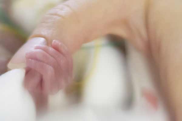
In chromosome 18 trisomy, various symptoms are caused by a chromosomal abnormality called trisomy, in which three chromosomes 18 are present. The main symptoms include short stature at birth, congenital heart disease, and microcephaly. It is a severe chromosome abnormality, and the one-year survival rate is considered to be about 10%. Recently, however, the prognosis of life has been gradually improving due to aggressive treatment such as neonatal intensive care and cardiac surgery.
It is estimated that Trisomy 18 occurs in 1 in 3,500 to 8,500 newborns.
Trisomy 18 causes a variety of symptoms due to a chromosomal abnormality called trisomy, in which three chromosomes 18 are present. The main symptoms include short stature at birth, congenital heart disease, and microcephaly. It is a severe chromosome abnormality, and the estimated one-year survival rate is about 10%. Recently, however, life expectancy has been gradually improving due to intensive newborn care, cardiac surgery, and other proactive treatments.
Many suffer from numerous complications, often resulting in spontaneous miscarriage or early death, but early detection through tests such as NIPT (New Non-invasive Prenatal Testing) allows proactive postnatal treatment and support.
The following is a detailed explanation of its symptoms, causes, testing methods, and prognosis.
What is Trisomy 18 (Edwards Syndrome)?
Trisomy 18 is a chromosomal disorder in which there is one extra chromosome 18.
This syndrome is named Edwards’ Syndrome after it was first reported in 1960 by John H. Edwards et al. in the United Kingdom. It is a serious disease, with most cases of fertilization not leading to successful delivery, most babies dying within a week of birth, and less than 10% surviving to one year of age, although there are some who live well past the age of 10 or 20 years.
There are 23 chromosomes in the human body, all of which are usually paired in pairs.
Trisomy is a condition in which the numbers of chromosomes are increased by one to three for some cause, and is thought to lead to a variety of conditions.
Some of the most common are the Down syndrome (trisomy 21) and trisomy 13 (Patau syndrome).
Occurrence Probability of Trisomy 18 by Gender
Trisomy 18 is a relatively rare disorder, and the current incidence of trisomy 18 (Edwards syndrome) is 1 in 3,500 to 8,500 newborns, with a gender ratio of Male:Female = 1:3, with a higher incidence in girls.
It is the second most common chromosomal abnormality in childbirth after Down syndrome (Trisomy 21), accounting for about 10% of reported cases. Because it is a severe chromosomal abnormality, miscarriages and stillbirths are relatively common during pregnancy.
Causes of Trisomy 18
In this section, we will explain how the chromosomal abnormality called trisomy 18 occurs and elaborate on its causes, explaining the basic biology of the syndrome.
What are chromosomes?
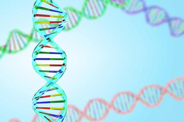
Chromosomes are rod-shaped structures found in the nucleus of cells. Chromosomes contain folded strands of DNA, which contain genetic information, and each chromosome contains multiple genes. There are 46 chromosomes in a human cell, 23 pairs of two chromosomes, one inherited from the father and the other from the mother. Chromosomes are generally numbered in order from the largest to the smallest (called “autosomes”), with chromosomes 1, then 2… up to chromosome 22. The cause of trisomy 18 is when the 18th autosome, chromosome 18, has three chromosomes instead of two.
Somatic Cell Division and Meiosis
So why do we have three chromosomes instead of two?
To understand the cause, we need to understand the mechanism of human cell division.
There are two major types of mechanisms by which human cells divide and multiply. One is somatic cell division and the other is meiosis.
Somatic cell division is a mechanism by which most cells in our body, such as skin, divide and multiply. When cells divide, the number of chromosomes doubles and divides into two, resulting in the same number of chromosomes (23 vs. 46) before and after cell division.
Meiosis, on the other hand, is a special cell division mechanism that occurs during the formation of reproductive cells such as sperm and egg. During meiosis, chromosomes are reduced, as the name “meiotic” division implies, one of the two paired chromosomes enters the sperm or egg. Thus, the resulting sperm or egg has 23 chromosomes, half of the original 23 pairs of 46 chromosomes. The meiosis-produced sperm and egg each have 23 chromosomes, and fertilization will result in 46 chromosomes. Thus, the fertilized egg of a child can inherit one chromosome each from the father’s sperm and the mother’s egg.
Chromosome Disjunction
However, during meiosis, for some reason, chromosome 18 may not divide properly and both chromosomes may end up in the sperm or egg. If this happens, an abnormal sperm or egg with two chromosome 18 is fertilized by a normal sperm or egg with one chromosome 18, resulting in a fertilized egg with three chromosome 18 (this is called ‘chromosome disjunction’).
It is believed that the majority of Trisomy 18 cases (80%) are caused by this chromosome disjunction. Although it is known that chromosome disjunctions are associated with old age at birth, the detailed mechanism has not yet been fully determined.
Trisomy 18 is caused by a genetic (chromosome) abnormality, but since the majority of cases are due to chromosome disjunction, it is thought to be caused by a ‘mutation’ rather than by a genetic problem in the parents that is inherited by the child.
About 10% are of the translocation type, in which a child may be born with trisomy 18 if part of chromosome 18 of one of the parents is bound to another chromosome (called a “translocation”). The parents may carry the translocated gene but have no symptoms and are carriers. In this type, the trisomy 18 is inherited from the parents to the child.
The last type, called mosaicism, is thought to account for about 10% of all cases. This type occurs when a fertilized egg is fertilized by a normal sperm and egg, and the fertilized egg undergoes cell division, resulting in the separation of three chromosomes (chromosome 18). This type is also not inherited from both parents, but is caused by mutation. In this type, normal cells and trisomy 18 cells are mixed together, so the symptoms are milder than in the other types.
Chromosome disjunctions are thought to be related to maternal age, and a 2015 RIKEN study and a Fujita Health University study found that this may be due to a decrease in factors that help maintain normal chromosome status as we age.
Below is a table showing the relationship between the mother’s age at childbirth and the incidence of trisomy 18 and trisomy 21.
Exhibit: Maternal Age and the Occurrence of Chromosomal Diseases at Birth,
Age of the Mother
(at birth)Occurrence Rate of
Trisomy 18Occurrence Rate of
Trisomy 2120 years old 1/10000 1/1441 25 years old 1/8300 1/1383 30 years old 1/7200 1/959 35 years old 1/3600 1/338 36 years old 1/2700 1/259 37 years old 1/2000 1/201 38 years old 1/1500 1/162 39 years old 1/1000 1/113 40 years old 1/740 1/84 41 years old 1/530 1/69 42 years old 1/400 1/52 43 years old 1/310 1/37 44 years old 1/250 1/38
Department of Obstetrics and Gynecology, Showa University School of Medicine, Tokyo, Japan.
Symptoms of Trisomy 18
Prenatal Suspected Findings
Prenatal symptoms include weak fetal movements, excessive amniotic fluid, dwarf placenta, and single umbilical artery. Fetal stunting, congenital heart defects, and morphological abnormalities of the wrists and ankles are also seen, and if suspected during ultrasound examinations during pregnancy, tests such as amniotic fluid testing and NIPT may be performed to check for chromosomal abnormalities.
Characteristics & Symptoms
The following physical characteristics and congenital diseases/complications are common in trisomy 18.
Cardiac diseases such as ventricular septal defects, atrial septal defects, and ductus arteriosus stenosis occur in 90% of patients and have a significant impact on prognosis, so early detection and treatment is important.
Congenital heart disease is considered to be a very frequent complication, occurring in approximately 90% of cases. Congenital heart disease is thought to affect survival rates due to the burden placed on the heart and the early development of congestive heart failure and pulmonary hypertension. Physical characteristics and the most frequent congenital heart diseases are as follows:
| Physical Characteristics | Potential Diseases and Complications |
|---|---|
|
|
Trisomy 18 Testing
Prenatal Testing
Quattro test: A prenatal blood test called the “Quattro test” measures four markers in the mother’s blood: beta-hCG, unconjugated estriol, inhibin, and alpha-fetoprotein.
How is Trisomy 18 (Edwards Syndrome) tested?
The examination for 18 trisomy is performed by ultrasound (echography),NIPT (New Non-invasive Prenatal Testing), etc.
If the baby is found to have any of the following morphological or chromosomal abnormalities in these tests, an amniocentesis or chorionic villus sampling will be performed to determine the number of chromosome 18 for a definitive diagnosis.
| Conditions in which trisomy 18 is suspected |
|---|
|
Treatments for Trisomy 18
There is no special treatment for trisomy 18, and more than half of the patients die within the first week after being born, and it is believed that less than 10% survive to the first year of life. In recent years, however, life expectancy has gradually improved with the aggressive use of neonatal intensive care, cardiac surgery, and esophagus-closure surgery.
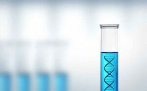
NIPT (Non-invasive Prenatal Testing)
A new type of prenatal genetic testing (NIPT: Non-invasive prenatal genetic testing) is also being used increasingly nowadays. Like the quattro test, this test requires blood samples from the mother, but it is a new screening test that analyzes the baby’s DNA fragments in the mother’s blood to check for chromosomal disorders and anomalies. It has a sensitivity of 99.9% and specificity of 99.6%, which is much more accurate in diagnosis than the Quattro test. Hiro Clinic NIPT performs this NIPT.
However, none of these tests can definitively confirm the diagnosis; they are only screening tests, and a definitive diagnosis requires an amniotic fluid test, which will be covered in a later section.
Trisomy 18 (Edwards syndrome)
and NIPT (New Non-invasive Prenatal Testing)
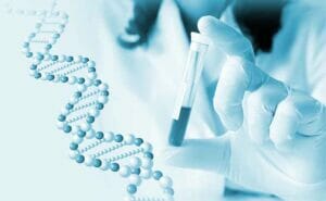
One of the tests used to detect abnormalities in babies before amniotic fluid and chorionic villus tests are performed is NIPT (New Non-invasive Prenatal Testing).
Blood is collected from the mother to examine fetal DNA in the blood to detect abnormalities.
Because NIPT (New Non-invasive Prenatal Testing) can be performed only with a blood test that does not affect the fetus, such as a miscarriage, and has little physical burden or risk to the pregnant woman, many people choose to undergo NIPT.
However, even if the physical risks are low, this is not a test that can be taken casually, and those who wish to undergo NIPT should read the following information carefully and make sure that your family understands the importance of the test.
This is the most important thing for your baby and your family.
Amniotic Fluid Testing
If trisomy 18 is suspected through a blood-based test, an amniotic fluid test is performed to confirm the diagnosis. A thin needle is inserted into the amniotic fluid to collect amniotic fluid while the condition of the fetus and amniotic fluid is monitored by ultrasound. The chromosomes of fetal cells contained in the amniotic fluid will be examined in detail.
NIPT (New Non-invasive Prenatal Testing) is a non-definitive test.
NIPT has the advantage of being more accurate than conventional maternal serum markers and ultrasound, but it is not always 100% accurate, with a higher positive result rate for mother with older age and a lower result rate for mother with younger age.
This is only a non-definitive test, and a positive result does not necessarily mean that the baby has a genetic abnormality.
An accurate diagnosis can only be made through definitive tests such as amniotic fluid and chorionic villus examinations.
Choose a Reliable Facility where you can get Counseling
The number of clinics offering NIPT has been increasing in recent years because the test is relatively easy to perform.
In response to this development, an increasing number of mothers are choosing to undergo NIPT without sufficient understanding of the details of the test.
It is reported that approximately 3% of all babies are born with some kind of congenital abnormality.
There is a possibility that unexpected difficult-to-treat illnesses may be found.
Some families undergo the test without understanding the purpose and content of the test and knowing that it is a non-definitive test, and upon receiving a positive result, they may become confused, disheartened, and unable to calmly make any further decisions.
NIPT is a way to plan and prepare in advance how the family will welcome the baby after birth and at which hospital the baby will receive appropriate treatment and support if abnormalities are found in the baby.
It is never intended to eliminate the life of the baby.
If you are considering NIPT, choose a clinic that has solid knowledge and experience and can provide counseling in advance.
NIPT is staffed by a specialist from the Japan Society of Obstetrics and Gynecology, a prenatal consulting pediatrician, and a clinical genetic specialist, so more specialized counseling is available.
It is also important to check in advance which clinics have pediatric specialists who can provide prompt care and treatment for the baby after diagnosis, and which clinics can work closely with psychiatrists and counselors who can provide emotional support for the mother and family.
【References】
- Japan Society of Obstetricians and Gynecologists – 染色体異常
- Information Center for Chronic Diseases of Childhood – MSD Manual Professional Edition – 18トリソミー
- Japan Society of Ultrasound Medicine – 産婦人科:胎児異常・胎児評価
- Ethics Committee of the Japan Society of Obstetrics and Gynecology – 母体血を用いた出生前遺伝学的検査(NIPT)に関する指針
We will look at the causes, symptoms and testing for 18 Trisomy (Edwards Syndrome) in detail / and how to prepare and prepare the family after the diagnosis.
Read more about NIPT
日本皮膚科学会 皮膚科専門医/日本医師会 産業医/東京衛生検査所 指導監督医
この記事は、 ヒロクリニックNIPTの編集・監修体制 にもとづき、資格を持つ医師が内容を確認しています。