

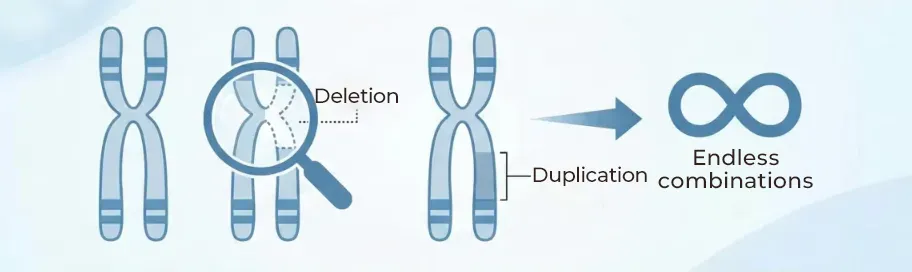
NIPT is used to screen for chromosomal abnormalities such as the following:
Primary Examples
Sex Chromosome Aneuploidies: Males typically have XY chromosomes and females XX, but patterns such as XXY, XYY, XXX, or a single X chromosome result in specific conditions.
Down Syndrome (Trisomy 21): A condition where there are three copies of chromosome 21.
Edwards Syndrome (Trisomy 18): A condition where there are three copies of chromosome 18.
Patau Syndrome (Trisomy 13): A condition where there are three copies of chromosome 13.
Partial Chromosomal Deletions or Duplications: Missing or extra pieces of a chromosome, which are almost always accompanied by intellectual and developmental disabilities after birth.
Among the congenital disorders caused by variations in the number of the 46 human chromosomes, the most frequent are Trisomy 21, sex chromosome numerical abnormalities, and partial deletions or duplications. Furthermore, Hiro Clinic performs a wide range of additional screenings, so let us take a closer look at various examples of these disorders on this page.

Basics of NIPT (Non-Invasive Prenatal Testing)
This is where the topic becomes a bit more technical.
Humans normally have 46 chromosomes, arranged into autosomes (22 pairs) and sex chromosomes (XY for males, XX for females). However, variations in the number or structure of these chromosomes can sometimes occur. For example, there are cases where there are three copies of chromosome 21 instead of the usual two. This is known as Trisomy 21 (Down syndrome), which brings the total number of chromosomes to 47.
When an excess or deficiency in the amount of chromosomal material occurs, it leads to an imbalance in the genetic data. This can result in congenital disorders or specific physical traits (chromosomal conditions), which significantly affect the baby’s development.
When people think of these conditions, they might rarely hear about any examples other than Down syndrome. Although other conditions do not receive much media coverage, it is a fact that sex chromosome abnormalities as well as disorders involving missing or extra pieces of genetic material (deletion and duplication disorders) are more common than most people think. Below are some representative disorders along with their frequencies.
In Japan, where the number of advanced maternal age pregnancies is on the rise, “Non-Invasive Prenatal Testing” (NIPT) is currently attracting significant attention. However, your honest reaction might be, “I don’t really know what kind of test it is or when I can take it.” Today, we bring you the knowledge to address those exact uncertainties.

There is still time for those up to their 15th week of pregnancy
About the NIPT costs you might be wondering about
Regarding Chromosomal Abnormalities and Their Frequency of Occurrence
In prenatal diagnosis, screening is conducted for various types of chromosomal abnormalities. These abnormalities can be broadly classified into the following two categories:
- Disorders with a different number of chromosomes: A condition where there is an anomaly in the “number” of chromosomes (e.g., trisomy or monosomy).
- Disorders with partial chromosomal deletions or duplications: A condition where a “piece of a chromosome is missing (deleted) or extra (duplicated).”
Below is a summary of representative chromosomal abnormalities and their frequencies of occurrence, listed in descending order of frequency.
| Rank | Condition Name | Abnormality Type | Frequency (Live Births) | Other Notes |
|---|---|---|---|---|
| 1 | Klinefelter Syndrome | XXY (Sex Chromosome) | Approx. 1/500 | Infertility or developmental characteristics seen in males. Sperm can be retrieved by performing TESE by age 30. |
| 2 | Triple X Syndrome | XXX (Sex Chromosome) | Approx. 1/800 | Mostly mild; symptoms may be unnoticeable in some cases. |
| 3 | Down Syndrome | Trisomy 21 | Approx. 1/1,000 | The most frequent chromosomal abnormality; a positive predictive value of 1/135 is documented. |
| 4 | Turner Syndrome | XO (Sex Chromosome) | Approx. 1/1,000 | Short stature, infertility, etc., seen in females. Early treatment is required from around age 3. |
| 5 | DiGeorge Syndrome | Microdeletion | Approx. 1/2,000 | Cardiac malformations, immune abnormalities, developmental disorders, etc. |
| 6 | Edwards Syndrome | Trisomy 18 | Approx. 1/3,500 | Mostly accompanied by severe disabilities, with high mortality rates under 1 year of age. A positive predictive value of 1/2,147 is documented. |
| 7 | 1p36 Deletion Syndrome | Microdeletion | Approx. 1/5,000 | Severe intellectual disability, seizures, hypotonia (low muscle tone), etc. |
| 8 | Patau Syndrome | Trisomy 13 | Approx. 1/5,000 | Severe malformations and central nervous system disorders; positive predictive value of 1/679. |
| 9 | Smith-Magenis Syndrome | Microdeletion | Approx. 1/15,000 | Self-injurious behavior, sleep disorders, developmental disorders, etc. |
| 10 | Wolf-Hirschhorn Syndrome | Microdeletion | Approx. 1/50,000 | Distinct facial features, developmental delays, seizures, etc. |
* Ordered by frequency of occurrence
Supplementary Note on Frequency of Occurrence
"Frequency of occurrence among live births" refers to the proportion among babies who are actually born.
What Can Be Understood from This Table
In addition to the well-known Down syndrome (Trisomy 21), there are various other disorders such as sex chromosome abnormalities and microdeletion/duplication syndromes, and these types of disorders are actually detected more frequently than Down syndrome.
Although individual cases of partial chromosomal deletions or duplications are relatively rare on a case-by-case basis, they represent critical anomalies linked to intellectual and developmental disabilities. Because there are so many different types of these variations, their combined total is said to exceed the number of Down syndrome cases.
At Hiro Clinic NIPT, it is possible to screen for 143 different types of variations in microdeletions and duplications alone.
This is the breakdown of the 32,923 patients who underwent screening at our clinic. (As of April 2023)
| Chromosome 21 |
Chromosome 18 |
Chromosome 13 |
Sex Chromosomes |
Other Chromosomes |
Partial Deletions/Duplications |
|
|---|---|---|---|---|---|---|
| Number of Affected Cases |
237 | 149 | 47 | 134 | 177 | 125 |
| Age 35 or Older |
187 | 120 | 29 | 77 | 111 | 69 |
| Under Age 35 |
50 | 29 | 18 | 57 | 66 | 56 |
| Ratio Between Age Groups |
3.74x | 4.14x | 1.61x | 1.35x | 1.68x | 1.23x |
Since all information is managed within our database at our clinic, we have accumulated over 60,000 cases of data to date and can provide relevant information as needed.
Description of Each Condition Detected by NIPT (Non-Invasive Prenatal Testing)
Trisomies 21, 18, and 13 (Accredited Facility Screening)
Trisomy 21 (Down Syndrome)
Among congenital disorders caused by variations in the number of the 46 human chromosomes, “Trisomy 21,” “Trisomy 18,” and “Trisomy 13” occur with the highest frequency.
The characteristics of these three conditions are as follows:
Trisomy 21 (Down Syndrome)
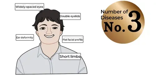
This condition is caused by having an extra copy of chromosome 21, and is also known as Down syndrome. Down syndrome is the most common genetic cause of intellectual disability, and its physical characteristics include developmental delays, low muscle tone (hypotonia), and distinct facial features.
Associated complications can include thyroid disorders, ENT (ear, nose, and throat) issues, and ophthalmic diseases. Some individuals may also experience structural organ issues, such as congenital heart defects, which can require medical or surgical treatment. The average IQ is reported to be around 50.
Many of these children attend local schools with support classes or specialized special needs schools, and some go on to thrive in various fields such as sports and the arts.
The average life expectancy is 60 years, and in many cases, individuals transition into specialized care facilities when their parents are no longer able to provide care.
Bing Image Search for Trisomy 21
Trisomy 18 (Edwards Syndrome)
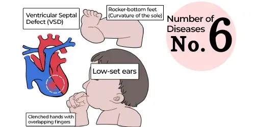
This condition is caused by having an extra copy of chromosome 18, and is also known as Edwards syndrome. Due to intrauterine growth restriction, most of these pregnancies result in miscarriage or stillbirth. Its physical characteristics include prenatal-onset growth failure, respiratory problems, and feeding difficulties.
Associated complications include heart defects (90%), gastrointestinal malformations, cleft lip and palate, and joint contractures. Even if born alive, most babies present with multiple congenital defects in the brain, heart, and other organs, resulting in high mortality rates under 1 year of age. In cases involving chromosomal mosaicism where children survive longer, they experience severe intellectual and developmental disabilities.
Bing Image Search for Trisomy 18
Trisomy 13 (Patau Syndrome)
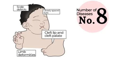
This condition occurs when there are three copies of chromosome 13 instead of two, and is also known as Patau syndrome. The vast majority of these children present with congenital defects in the brain and other vital organs.
Its physical characteristics include growth failure, respiratory problems, and feeding difficulties.
Associated complications include cleft lip and palate, polydactyly (extra fingers or toes), eye disorders, heart defects (80%), and holoprosencephaly, resulting in structural congenital anomalies in the brain and other organs.
Most pregnancies result in miscarriage or stillbirth. Even among those born alive, 80% pass away before reaching one month of age, and 10% do not survive past their first year, making it a condition with high early mortality. It is known that when a child is born with chromosomal mosaicism—where only a portion of the cells have three copies of chromosome 13 instead of all of them—the severity of the symptoms can be somewhat milder.
Bing Image Search for Trisomy 13
Sex Chromosome Numerical Abnormalities Detected by NIPT (Infertility Screening)
Sex chromosome disorders arise from sex chromosome aneuploidy, which is caused by nondisjunction during cell division. It is said to be influenced by the age of both parents. Because this category includes a wide variety of conditions, its total number of occurrences actually exceeds that of Down syndrome when looking at total live births. Although the clinical symptoms are often described as relatively mild, they vary significantly on an individual basis and cannot be generalized.
Below are examples of conditions caused by numerical abnormalities of the sex chromosomes. Since there are various patterns, let’s look at each of them in detail.
Klinefelter Syndrome (47,XXY / 48,XXXY / 46,XY / 47,XXY [Mosaic])
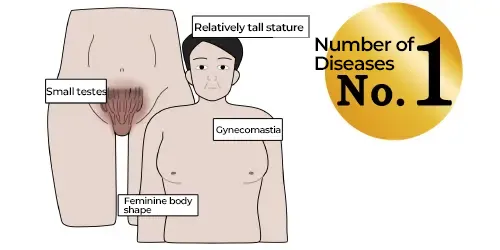
The frequency of occurrence is approximately 1 in 1,000 live male births.
In Klinefelter syndrome, about half of the cases are reported to be caused by non-disjunction of paternal chromosomes. Cases stemming from maternal non-disjunction are influenced by advanced maternal age at childbirth.
Because individuals have a typical male outward appearance, the condition often goes unnoticed during childhood. Symptoms include incomplete development during puberty (reduced body hair and muscle development), tendencies toward feminization (gynecomastia/breast development), larger hands and feet with long lower limbs (tall stature), and small testes resulting in azoospermia (infertility). There is rarely significant intellectual impairment or impact on general life expectancy, though cases with a higher number of extra X chromosomes may present with lower intellectual ability.
Bing Image Search for Klinefelter Syndrome
Turner Syndrome (45,X)
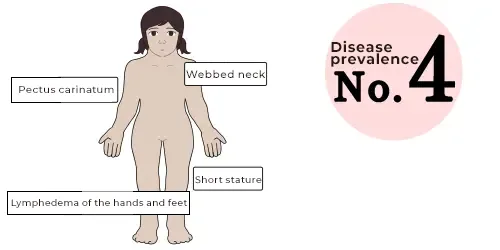
The frequency of occurrence is approximately 1 in 2,500 live female births.
Turner syndrome is not dependent on maternal age. Approximately 80% of Turner syndrome cases present as a monosomy (a single X chromosome). It is known that the remaining X chromosome is typically of maternal origin, meaning the paternal X chromosome is most often the one that was lost.
It is estimated that about 99% of X monosomy embryos result in spontaneous miscarriage.
Incidence in all pregnancies: At the fertilized egg stage, it is estimated to occur in about 3% of all pregnancies.
Probability of reaching live birth: Out of those, less than 1% successfully survive to live birth.
Proportion of total miscarriages: Data from chorionic villus sampling after spontaneous miscarriages show that X monosomy accounts for roughly 10% of those cases.
Outwardly, individuals have a female appearance. Symptoms include lymphedema, cubitus valgus (turned-out elbows), webbed neck, short stature, delayed sexual maturation, and amenorrhea (absence of menstruation).
In most cases, it results in infertility.
Regarding intellectual disability, cases range from having no impairment at all to mild cognitive difficulties.
Bing Image Search for Turner Syndrome
Triple X Syndrome (47,XXX)
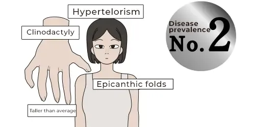
The frequency of occurrence is approximately 1 in 1,000 live female births.
Much like typically developing females, individuals with Triple X syndrome maintain only a single active X chromosome per cell. Even with three X chromosomes, two of them undergo X-inactivation (becoming genetically inactive), which is why there are generally no distinct physical abnormalities. Additionally, individuals typically have normal fertility.
Bing Image Search for Triple X Syndrome
XYY Syndrome (47,XYY)
The frequency of occurrence is approximately 1 in 1,000 live births.
Overview and Characteristics of 47,XYY (Jacobs Syndrome)
1. Physical Characteristics
Height: Growth velocity is accelerated starting from early childhood, with the final average adult height being approximately 7 cm taller than predicted. (For example, a study in Scotland showed that for fathers averaging 174 cm and mothers averaging 162 cm, the average height of their XYY sons reached 188 cm).
Cause: The increased height is believed to be caused by having an extra copy of the “SHOX gene,” which is located on the X and Y chromosomes.
Sexual Development and Fertility: Prenatal testosterone levels are normal. Most males experience typical sexual development and have normal fertility.
2. Intelligence and Learning (Cognitive and Behavioral Profile)
IQ (Intelligence Quotient): Unlike other sex chromosome abnormalities (such as Klinefelter syndrome), the average IQ of boys with XYY is not reduced compared to the general population.
The average Full Scale IQ across multiple studies is around “105” (within the average range).
However, when compared directly to their siblings, their scores tend to be slightly lower.
Learning: Approximately half of these children may experience learning difficulties, particularly in reading and writing. While this is a higher rate than seen in children with typical IQs, it can be well-managed with appropriate academic support and remedial classes.
3. Behavior and Development
Developmental Delays and Behavior: While developmental delays or behavioral characteristics can sometimes be observed, individual variation is immense, and these are not symptoms unique to XYY. Management and support strategies are identical to those used for typical boys (46,XY).
Aggression: Contrary to outdated theories, the claim that “males with XYY show higher levels of aggression” has been scientifically disproven.
Psychological Aspects: Some psychological studies suggest there may be challenges regarding impulse control and emotional regulation.
4. Other Medical Risks
Complications: A slightly higher risk for asthma, seizures, and tremors has been reported.
Urological System: In very rare cases, hypoplasia of the testes or penis may be present, which can cause infertility due to reduced sperm count or abnormalities.
5. Cause of Occurrence (Genetics)
Not Inherited: XYY is not inherited from parents; it is a random event caused by an accidental chromosomal “nondisjunction” during sperm production (meiosis II).
Mosaicism: If nondisjunction occurs during the early stages of cell division after fertilization, it can result in a “mosaic” state where typical cells (46,XY) and XYY cells coexist.
Bing Image Search for XYY Syndrome
Fetal Gender Determination
Fetal gender is determined by the sex chromosome carried by the father’s sperm.
Consequently, the gender is established at the exact moment of fertilization; if a sperm carrying a Y chromosome fertilizes the mother’s egg, the baby will be a boy, and if a sperm carrying an X chromosome fertilizes the egg, the baby will be a girl. While it is standard to identify fetal gender via ultrasound starting around 18 to 20 weeks of pregnancy, NIPT can determine gender as soon as the pregnancy is confirmed by an ultrasound scan.
The most medically significant indication for identifying fetal gender is for families with a history of X-linked recessive genetic disorders. For instance, if the fetus’s grandfather has an X-linked recessive condition, his daughter is typically a carrier (possessing the altered gene without showing symptoms). If she becomes pregnant and undergoes fetal gender determination, it can be deduced that a male fetus has a 50% chance of developing the condition, while a female fetus has a 50% chance of becoming a carrier. Naturally, the remaining 50% of male fetuses and 50% of female fetuses will be genetically unaffected, meaning the gene will not be passed down to future generations.
NIPT screenings that analyze sex chromosomes are not performed at accredited facilities in Japan. However, because sex chromosome abnormalities occur with relatively high frequency, it is widely recommended in the United States and Europe that pregnant individuals undergo this screening.
NIPT (Non-Invasive Prenatal Testing) Deletion and Duplication Syndromes (Intellectual Disability Screening)
This screening detects whether specific regions of a chromosome are missing (deletion) or extra (duplication). When screening was conducted on 10,000 pregnant individuals, it was found that approximately 1 in 285 showed a chromosomal deletion or duplication. It is well established that the extent of genetic material altered (gained or lost) correlates with the severity of the clinical symptoms. When a deletion or duplication spans 7 million base pairs (7 Mb) or more, there is a high probability that the child will be born with intellectual or developmental disabilities, even if the clinical severity does not reach that of a full trisomy or monosomy. For example, while a fetus with an entire extra copy of Chromosome 1 cannot survive, survival is highly possible if the anomaly is a partial deletion or duplication of Chromosome 1. At Hiro Clinic NIPT, we consider these to be conditions where survival is viable, though accompanied by developmental or health challenges.
Important Note: While it is possible to screen for deletions or duplications in major regions of autosomes, these variations cannot be detected if the affected region is smaller than 7 million base pairs (7 Mb).
Below are some of the representative syndromes associated with chromosomal deletions and duplications.
143 Types of Microdeletion and Duplication Syndromes
143 Types of Microdeletion and Duplication Syndromes (Intellectual Disability Screening)
1p36 Deletion Syndrome
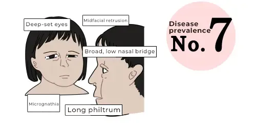
1p36 deletion syndrome is a congenital disorder caused by a microdeletion in a specific region on the short arm (p) of chromosome 1.
Symptoms include distinctive facial features, developmental and intellectual delays, and epileptic seizures (convulsions, impaired consciousness). Additionally, individuals may be born with congenital heart defects.
Bing Image Search for 1p36 Deletion Syndrome
4p Deletion Syndrome (Wolf-Hirschhorn Syndrome)
4p deletion syndrome is also known as “Wolf-Hirschhorn syndrome” and is a congenital disorder caused by a deletion in a specific region on the short arm (p) of chromosome 4.
Characteristics include severe intellectual and developmental delays, growth failure, intractable epilepsy (seizures), and multiple structural malformations.
Bing Image Search for 4p Deletion Syndrome
Chromosome 17 Microdeletion: Smith-Magenis Syndrome
Smith-Magenis syndrome is a congenital anomaly syndrome that affects many parts of the body. Its primary characteristics include mild to moderate intellectual disability, delayed speech and language skills, distinctive facial features, sleep disturbances, and behavioral problems. Smith-Magenis syndrome is caused by a microdeletion of genetic material from chromosome 17 in each cell. This deletion occurs on the short (p) arm of the chromosome at a position designated as p11.2.
Bing Image Search for Smith-Magenis Syndrome
22q11.2 Deletion Syndrome (DiGeorge Syndrome)
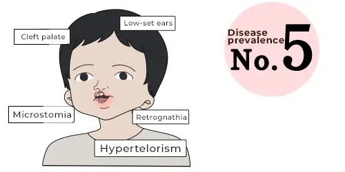
Also known as “DiGeorge syndrome,” this is a congenital disorder caused by a microdeletion of a specific region on chromosome 22, resulting in the loss of approximately 30 genes.
Symptoms include congenital heart defects, intellectual and developmental delays, distinctive facial features, immune system deficiencies, cleft palate or velopharyngeal insufficiency, a nasal voice, and hypocalcemia (low blood calcium levels).
Bing Image Search for DiGeorge Syndrome

NIPT (Non-Invasive Prenatal Testing) Full Chromosome Aneuploidy Screening (Miscarriage Risk Screening)
At Hiro Clinic NIPT, we offer “Full Chromosome” screening (chromosomes 1 to 22, plus X and Y), extending beyond just trisomies 21, 18, and 13. While full trisomies in every cell often present severe survival challenges, individuals with “mosaicism”—where a mixture of typical and atypical cells exists—have been documented to survive across all chromosome pairs from 1 to 22. The incidence of these chromosomal variations shows an upward trend with advanced maternal age. Our screening detects both trisomies (extra chromosomes) and monosomies (missing chromosomes).
This comprehensive test is highly recommended for pregnant individuals aged 35 and older. It is a standard screening practice in countries with advanced prenatal care models, such as Belgium.
Our Clinic’s Sensitivity and Specificity
Autosomal Trisomies
| Condition | Sensitivity [95% CI] |
Specificity [95% CI] |
Positive Predictive Value (PPV) [95% CI] |
|---|---|---|---|
| Trisomy 21 | >99.9% (98.1–100.0%) |
99.9% (99.9–100.0%) |
94.4% (90.5–96.8%) |
| Trisomy 18 | >99.9% (93.7–100.0%) |
>99.9% (99.9–100.0%) |
85.1% (74.7–91.7%) |
| Trisomy 13 | >99.9% (81.6–100.0%) |
>99.9% (99.9–100.0%) |
65.4% (46.2–80.6%) |
Sex Chromosome Aneuploidies
| Condition | Sensitivity [95% CI] |
Specificity [95% CI] |
Positive Predictive Value (PPV) [95% CI] |
|---|---|---|---|
| XO | >99.9% (79.6–100.0%) |
99.6% (99.5–99.7%) |
30.0% (19.1–43.8%) |
| XXX | >99.9% (77.2–100.0%) |
99.9% (99.8–100.0%) |
59.1% (38.7–76.7%) |
| XXY | >99.9% (75.8–100.0%) |
99.9% (99.8–99.9%) |
48.0% (30.0–66.5%) |
| XYY | >99.9% (51.0–100.0%) |
99.9% (99.9–100.0%) |
40.0% (16.8–68.7%) |
Partial (Micro) Deletions / Duplications
| Condition | Sensitivity [95% CI] |
Specificity [95% CI] |
Positive Predictive Value (PPV) [95% CI] |
|---|---|---|---|
| Partial (Micro) Deletions / Duplications | >99.9% (90.1–100.0%) |
99.1% (99.0–99.2%) |
16.4% (12.1–22.0%) |