

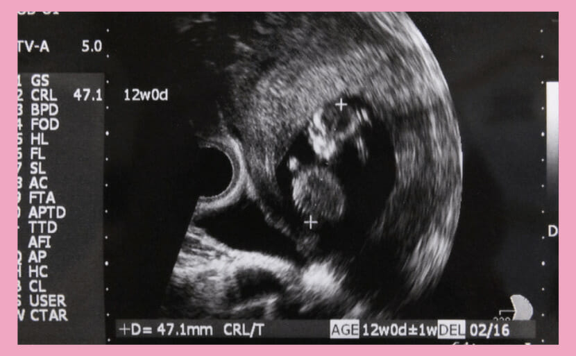
Follow the poignant journey of a mother and baby at 12 weeks as Down syndrome is revealed by an echocardiogram, with sensitive insights supervised by our medical experts at Hiro Clinic.

List of Test Plans
for Down Syndrome
Summary of this article
By 12 weeks of pregnancy, an ultrasound scan can already pick up findings (such as nuchal translucency, or NT) that are associated with Down syndrome. Even so, seeing such a finding does not confirm that the baby has Down syndrome. An ultrasound is a screening tool that estimates likelihood only – a definitive diagnosis requires a diagnostic test such as chorionic villus sampling (CVS) or amniocentesis. This article looks at week 12 from an obstetrician’s perspective: how the baby and mother are changing, what an ultrasound can and cannot tell you, and how these findings connect to NIPT (Non-Invasive Prenatal Testing).
What this article covers
- How big your baby is at 12 weeks, and whether the sex can be determined
- What a 12-week ultrasound can show, and when Down syndrome-related findings become visible
- How to correctly read (and not over-read) findings such as NT (nuchal translucency)
- How a finding connects to NIPT and diagnostic testing
- How your body is changing at 12 weeks, and what to watch for

Your Baby at 12 Weeks: Size, Weight, and Sex
By 12 weeks, your baby’s organs have largely finished forming, and the body is starting to look distinctly human. Let’s look at the typical size and weight, and whether the sex can be told yet.
Size and Weight at 12 Weeks
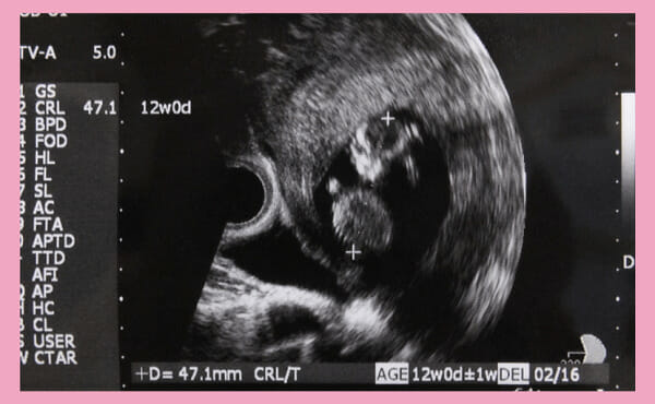
At around 12 weeks, your baby’s crown-rump length (CRL) – the measurement from the top of the head to the buttocks – is about 5-6 cm. That’s roughly the size of a small mandarin orange, and the baby weighs about 40-50 g, close to the weight of an egg.

Physically, the major organs have essentially finished forming, and facial features and muscles are gradually becoming more defined. The volume of amniotic fluid is increasing too, so your baby’s arms and legs move more actively. Differences in the external genitalia are starting to appear, so the body is beginning to look distinctly human.
Can You Tell the Baby’s Sex at 12 Weeks?
Around 12 weeks, differences in the external genitalia begin to appear, so an ultrasound can sometimes reveal the baby’s sex. That said, growth at this stage can still be too limited to make a confident call, so it isn’t always possible to know for certain.
NIPT analyzes the baby’s DNA from a sample of the mother’s blood, so it can determine sex-related information with high accuracy as well. That said, facilities accredited by the Japan Society of Obstetrics and Gynecology do not perform NIPT for the sole purpose of sex determination – it is only ever an incidental finding within a test that primarily screens for chromosomal conditions.
What a 12-Week Ultrasound Can Show, and When Down Syndrome Findings Become Visible
By 12 weeks, you’re inside the window in which ultrasound findings associated with Down syndrome (such as NT) can be observed. That window opens at 11 weeks 0 days and runs through 13 weeks 6 days, so week 12 falls right in the middle of it. But seeing a finding is not the same as a diagnosis. Let’s walk through this stage by stage so the two don’t get confused.
On ultrasound, doctors assess the likelihood of trisomy 21 (Down syndrome), a well-known chromosomal condition, by looking at partial features such as the back of the baby’s neck and the length of the limbs. Even if the length of the limbs or the size of the face falls outside the average range, that alone does not confirm trisomy 21 – it only raises a suspicion. Some pregnancies go undetected until birth, so try not to draw firm conclusions from a week-12 finding alone.
| Stage of pregnancy | Main purpose of ultrasound | Main findings related to Down syndrome |
|---|---|---|
| Up to week 10 | Confirming the gestational sac, heartbeat, and dating | No findings can be assessed yet |
| Weeks 11d0-13d6 (includes week 12) | Combined test / early detailed ultrasound | Increased NT (nuchal translucency), absent/underdeveloped nasal bone, tricuspid regurgitation, shortened femur (FL) |
| Weeks 18-22 (mid-pregnancy) | Fetal anatomy screening | Heart defects, duodenal atresia, shortened femur, cleft lip/palate, and others |
| Diagnostic testing | Definitive chromosome diagnosis | CVS (around weeks 11-14) / amniocentesis (from around weeks 15-16) |
The key point is that every one of these is simply a “soft marker” – a sign that raises probability, not a proof. Plenty of babies with a finding turn out to have typical chromosomes, and some babies with Down syndrome show no findings at all. Treat an ultrasound finding as one piece of evidence for estimating likelihood, not a verdict.
Nuchal Translucency (NT)
The finding given the most weight on a 12-week ultrasound is fluid buildup in the soft tissue behind the baby’s neck. Clinically it’s called “NT” (Nuchal Translucency), and it shows up as a dark, translucent band on the scan image.
This fluid is a physiological feature seen in every baby. Because the circulatory system is still immature, it tends to stand out temporarily around 11-13 weeks and, in most cases, disappears naturally once circulation matures around weeks 16-18. That’s exactly why the timing and technique of the measurement matter so much for the result.
NT is measured at the thickest point of the mid-sagittal plane (the plane that divides the body evenly left and right), when the CRL (crown-rump length) is between 45 and 84 mm. As a rough guide, values above 3.0-3.5 mm are generally flagged for further evaluation, but the reference threshold varies by CRL and by clinic, so this number alone doesn’t confirm anything.
The measurement itself is sensitive to the examiner’s skill. NT reads thicker when the baby’s neck is extended and thinner when it’s flexed, so an accurate measurement takes real technique. Larger NT values are associated with a higher rate of chromosomal or cardiac abnormalities, but many babies with an above-threshold NT turn out to have entirely typical chromosomes.
Here’s what an actual mother who was told about an ultrasound finding shared. In my own clinic, when I tell a patient “the nuchal area looks a little thicker than average,” I regularly see the anxiety cross their face. On X (formerly Twitter), a mother posted about being told at 12 weeks 6 days that her baby had a fluid buildup around the neck (a cystic hygroma); @momomooss wrote about how shaken she felt at the possibility of a chromosomal condition. It’s completely natural to feel unsettled when a finding turns up. What matters most is understanding what the finding actually means, and then talking with your doctor about how to move forward with testing.
Limb Length (FL)
Chromosomal conditions such as trisomy 21 are sometimes associated with shorter limbs. Femur length (FL) is one of the measurements used as a supporting data point. That said, a shorter FL is often simply a slower growth pattern, so it can’t be judged on its own.
Head Size (BPD/FOD)
Doctors also look at trends in BPD (Biparietal Diameter, the width of the skull) and FOD (Front-Occipital Diameter, the front-to-back length). At 12-15 weeks, a BPD of roughly 15-35 mm is typical, and a value that departs significantly from average prompts further evaluation. As a rule, no single measurement is judged in isolation – it’s tracked alongside how the pregnancy progresses.
Nasal Bone and Facial Features
With trisomy 21, the facial profile is sometimes described as appearing generally flatter, and on ultrasound the nasal bone can appear small or hard to visualize. Nasal bone height becomes assessable once the CRL reaches 45-84 mm, which lines up closely with around week 12.
Heart (Tricuspid Regurgitation and More)
About half of babies with trisomy 21 have some form of heart condition. If blood flows backward across the tricuspid valve, between the right ventricle and right atrium, that raises suspicion of both congenital heart disease and trisomy 21. That said, regurgitation alone also shows up in babies without trisomy 21, so it doesn’t confirm anything by itself. Because the heart is difficult to fully assess in the first trimester (up to around week 15), a specialist typically evaluates it in more detail with a dedicated “fetal echocardiogram” before drawing any conclusion.
Does a Baby Moving Vigorously on Ultrasound Signal Down Syndrome?
Quite a few expectant mothers see their baby kicking and bouncing energetically during a scan and worry that all that movement might be a sign of Down syndrome.
The short answer: there’s no direct link between how vigorously a baby moves on ultrasound and chromosomal conditions such as trisomy 21. Around 12 weeks, muscles and nerves in the limbs are developing rapidly, so it’s common to see your baby kicking the amniotic membrane or waving its arms and legs around in the fluid. That’s simply solid evidence that your baby is growing well and developing normal motor function.
Suspicion of Down syndrome is based on structural findings – NT and the measurements covered above – not on how active your baby looks. A baby moving energetically is not, in itself, something to worry about, so try to relax during your scan.
If a Finding Comes Up: How NIPT and Diagnostic Testing Connect
If your ultrasound turns up a finding, the next step is understanding the difference between “screening” and “diagnostic” testing. Ultrasound and NIPT are screening tests that estimate probability; CVS and amniocentesis are diagnostic tests that provide a definitive answer. Think of it as a two-stage process.
What Is NIPT?
NIPT (Non-Invasive Prenatal Testing) analyzes cell-free DNA (cfDNA) from the baby that circulates in the mother’s blood, to assess the likelihood of certain chromosomal conditions. It’s reported to be highly accurate for specific conditions such as trisomy 21 (Down syndrome), but it’s crucial to remember that it remains a screening test, not a diagnostic one. A negative result does not reduce the possibility to zero, and a positive result still needs to be confirmed with CVS or amniocentesis.
Real expectant mothers around week 12 have shared their thoughts on booking NIPT or a detailed fetal ultrasound. @newrysheep posted that, heading into a check-up just before hitting 12 weeks, she planned to “book NIPT and, if possible, a fetal ultrasound package too” once everything looked fine. It’s clear that many mothers think about ultrasound and NIPT together, as a combination. In my own clinic I tell patients the same thing – ultrasound and NIPT serve different roles, so combine them as needed. Some mothers specifically want a more detailed ultrasound; @pojipojiyuri posted that she preferred “a facility that can also do a detailed early ultrasound at the same time,” which reflects a shared desire to have early findings looked at thoroughly.
Timing: CVS vs. Amniocentesis
There are two types of diagnostic test – CVS and amniocentesis – and they’re available at different times. Here’s how they compare.
| Test | Typical timing | Key features |
|---|---|---|
| Chorionic villus sampling (CVS) | Around weeks 11-14 | Samples tissue from the placenta. Provides a definitive diagnosis and can be done relatively early |
| Amniocentesis | From around weeks 15-16 | Samples amniotic fluid. Provides a definitive diagnosis, with a slightly lower miscarriage risk than CVS |
NIPT only requires a blood draw from the mother’s arm, so it carries no risk of miscarriage. It is not covered by health insurance in Japan, and the self-pay cost generally runs about 90,000-240,000 yen depending on the test scope. Because prenatal testing is not a treatment, it also does not qualify for the Japanese medical expense tax deduction.
Timing matters too. Because amniocentesis, a diagnostic test, isn’t available until around weeks 15-16, NIPT needs to be completed – and its results returned – before then. At Hiro Clinic NIPT, testing is available from early on, right after the heartbeat is confirmed by ultrasound, and there is no upper age limit. Samples are analyzed in Japan, in partnership with a domestic testing institution (the Tokyo Health Inspection Center), so results are typically delivered in as little as 2 to 5 days.
Tests Available at 12 Weeks: Antenatal Checkups and Self-Pay Tests
Around 12 weeks, you’ll have your routine antenatal checkup, and you can also choose from several self-pay tests that screen for genetic and chromosomal conditions. Here’s what each involves.
Antenatal Checkups
An antenatal checkup is a regular examination throughout pregnancy that monitors the health of mother and baby. Under guidelines from Japan’s Ministry of Health, Labour and Welfare, checkups around week 12 are recommended roughly once every four weeks. The main items covered are:
- Basic measurements (height, weight, BMI, blood pressure, etc.)
- Blood tests
- Cervical cytology test
Let’s look at each of these in more detail.
Basic Measurements (Height, Weight, BMI, Blood Pressure, etc.)
Antenatal checkups start by gathering the basic information needed to manage your pregnancy. Your care team records height, weight, BMI, blood pressure, urine protein, and urine sugar, and uses a questionnaire to collect more detailed information from you.
Blood Tests
Your blood test checks the following:
- ABO blood type and Rh factor
This confirms your blood type in case a transfusion is needed, and checks for blood-type incompatibility between mother and baby that could trigger antibody formation.
- Irregular antibody screening
This checks your blood for irregular antibodies. If present, these antibodies can react with the baby’s blood cells and break them down.
- Complete blood count
This measures components such as white blood cells, red blood cells, hemoglobin, and platelets, revealing signs of inflammation, anemia, immune status, and clotting ability.
- Blood glucose
This checks the sugar level in your blood. During pregnancy, enzymes released by the placenta can make blood sugar harder to control, raising the risk of gestational diabetes.
- HBs antigen, HCV antibody, rubella antibody, syphilis screening, HTLV-1 antibody, HIV screening, toxoplasma antibody
These tests confirm whether you carry particular antigens or antibodies. Knowing your status makes it easier to reduce the risk of transmission to your baby.
Cervical Cytology Test
This test collects cells from the cervix and examines them under a microscope for abnormalities, helping detect cervical or vaginal cancer. In early pregnancy, this test is generally recommended for women who haven’t been screened within a certain recent period.

Self-Pay Tests Available From 12 Weeks
Beyond your routine antenatal checkups, there are self-pay tests for genetic and congenital conditions: the maternal serum marker test (also known as the Quad or Quattro test), the combined test, and NIPT.
The maternal serum marker test is done around weeks 15-17, the combined test around weeks 11-13, and NIPT anywhere from weeks 10-22. Because each of these tests has a fixed availability window, waiting too long to decide can mean you miss the chance to take the test you wanted. It’s worth starting your research early.
In Japan, the cost of NIPT for a basic panel covering trisomy 21, 18, and 13 typically runs about 90,000-240,000 yen (before tax), and prices vary between facilities. At Hiro Clinic NIPT, we offer plans tailored to the scope of testing you want – from a single-chromosome plan, to a plan covering chromosomes 21, 18, and 13, to broader plans for those who want to know about a wider range of possibilities.
One thing to keep in mind about NIPT: it doesn’t give you a simple “yes or no” answer. Because it’s a screening test, the result tells you whether the probability of a chromosomal condition is “high” or “low,” not whether the condition is definitely present or absent. The blood draw involves some minor discomfort, but it’s not considered to carry meaningful risk to the baby.
If you move on from NIPT to a diagnostic test such as CVS or amniocentesis, it’s worth knowing that these procedures do carry a small but real risk of miscarriage or premature rupture of membranes. That said, a diagnostic test is never mandatory just because a screening result is positive – it’s only done if and when you choose to move forward. We’re happy to walk you through plan details and important considerations, so please reach out with any questions.
How a Mother’s Body Changes at 12 Weeks
What kind of changes happen in a mother’s body around week 12? Here’s a look at the maternal side of things, and what to watch out for.
Maternal Changes
Around week 12, your belly starts to gently round out as your baby grows. This is also roughly when the placenta – which exchanges nutrients and oxygen with your baby and produces blood – finishes forming. For many women, morning sickness (loss of appetite and nausea) starts to ease noticeably around this point.
As morning sickness fades, appetite often comes roaring back, and it’s easy to overeat without noticing. Aim for a balanced diet to avoid sudden weight gain.
What to Watch For at 12 Weeks
By 12 weeks, your body is going through a number of changes. Here’s what’s worth keeping in mind.
Choose Clothing That Doesn’t Press on Your Belly
At 12 weeks, your belly may feel a little fuller or tighter. It’s not dramatically noticeable yet, but it’s best to avoid tight clothing that presses on your abdomen. Pressure can restrict blood circulation or trigger excessive uterine contractions, potentially putting stress on your baby. Choose roomier clothes or maternity wear that give your belly some breathing room.
More Frequent Urination
As your baby grows, added pressure on the bladder can make you feel the urge to urinate more often. Even if you’re heading to the bathroom more, don’t hold it in. Pregnancy hormones loosen the urethra, which makes it easier for bacteria to take hold if you hold your urine – so relieve yourself as soon as you feel the need.
Abdominal discomfort is also common at this stage. Many women notice some abdominal pain up to around week 15. It can stem from your baby’s growth and the changing size of the uterus, hormone-driven constipation, or diarrhea from shifting bowel function. If the discomfort gradually eases, it’s usually nothing serious, but early miscarriage remains a possibility before week 12. If the pain intensifies or you notice bleeding, talk to your doctor.

What Is Down Syndrome? (The Basics)
Down syndrome is a chromosomal condition caused by having an extra copy of chromosome 21. Its formal name is “Down syndrome (trisomy 21)”, and it’s the most common chromosomal condition among newborns.
The extra chromosome often affects muscle and organ development, and early support can be genuinely helpful for developmental delays. Distinctive facial features, hand shape, slower intellectual development, and associated heart conditions can occur, but the degree varies considerably from person to person. Down syndrome was once associated with a short life expectancy, but medical advances have extended the average life expectancy in Japan to roughly 60 years.
Down syndrome falls into three types: standard trisomy 21 (about 95%), translocation type (about 3%), and mosaic type (about 2%). Only a portion of the translocation type – roughly 1.5% of all Down syndrome cases – is clearly inherited from a parent. The overwhelming majority occur by chance, and are not caused by anything the parents did or didn’t do.
Down Syndrome Frequency by Maternal Age
Overall, a baby is born with Down syndrome in roughly 1 in 1,000 pregnancies (0.1%). The frequency rises with the mother’s age at delivery, exceeding 1% from age 40 onward. These figures are approximate guides only – published numbers vary somewhat between sources.
| Maternal age | Approximate frequency | Approximate rate |
|---|---|---|
| 20 | 1 in 1,667 | About 0.060% |
| 30 | 1 in 952 | About 0.105% |
| 35 | 1 in 378 | About 0.260% |
| 40 | 1 in 106 | About 0.943% |
| 45 | 1 in 30 | About 3.333% |
| 49 | 1 in 11 | About 9.091% |
Frequency rises with age mainly because egg aging increases the rate of nondisjunction – a failure of chromosomes to separate properly. If maternal age is a concern for you, combining ultrasound with NIPT can help you move through pregnancy with a clearer picture, rather than carrying uncertainty the whole way.
Things to Do at 12 Weeks
Once you hit 12 weeks, here’s what’s worth taking care of.
- Weight management
- Preparing maternity wear
- Stretch mark care
- Considering NIPT
For many women, morning sickness gradually eases around 12 weeks. Watch out, though – as your appetite picks back up, weight can climb quickly. Manage your intake to avoid rapid weight gain. Some women find their morning sickness lingers longer, which can stem from your baby’s growth pressing on internal organs, hormonal shifts, stress, or poor circulation. Since the cause differs from person to person, there’s no single fix that works for everyone – it helps to find your own way of coping.
By around week 12, unlike the very early weeks of pregnancy, your belly starts noticeably growing bigger by the day. As it expands, your existing clothes can start pressing uncomfortably on your abdomen, so it’s a good time to prepare maternity wear. As your belly grows, you may also develop stretch marks on your abdomen. Keeping the skin moisturized with creams or oils, along with gentle massage, can help prevent them.
If you’d like to learn about your baby’s health sooner rather than later, consider NIPT (Non-Invasive Prenatal Testing). Knowing more about your baby’s condition ahead of delivery can help you prepare at your own pace, with a calmer mind.
If a Finding Turns Up: Where to Get Support
If an ultrasound finding is followed up and testing suggests a high likelihood of Down syndrome, it’s important not to carry that weight alone – reach out to specialists. Genetic counseling can help you work through whether to pursue further testing and what comes next, together with a professional. Connecting with local parent support groups or organizations such as the Japan Down Syndrome Society that support individuals and families can also be a real source of comfort.
Frequently Asked Questions
Can a 12-week ultrasound detect Down syndrome?
At 12 weeks, you’re within the window where findings related to Down syndrome, such as NT, can be observed. But seeing a finding is not a diagnosis – it’s only an estimate of likelihood. Confirming a diagnosis requires CVS or amniocentesis.
If NT (nuchal translucency) is above the reference range, does that confirm Down syndrome?
No. Many babies with an above-range NT turn out to have entirely typical chromosomes. NT is just one piece of evidence used to estimate probability. If your NT value concerns you, talk with your doctor about NIPT or a diagnostic test.
Can you tell the baby’s sex at 12 weeks?
Because differences in the external genitalia start to appear around this time, ultrasound can sometimes reveal the sex, though it isn’t always possible depending on how much the baby has grown. NIPT also provides sex-related information, but accredited facilities do not perform the test solely to determine sex.
If an ultrasound finding comes up, what should I do next?
The typical path is to first look more closely at the likelihood with a screening test such as NIPT, and if that comes back positive, confirm the diagnosis with CVS or amniocentesis. Talk with your doctor about the right order and timing for your situation.
What tests are available at 12 weeks?
Beyond your routine antenatal checkup, self-pay options include the maternal serum marker test (around weeks 15-17), the combined test (around weeks 11-13), and NIPT (weeks 10-22). Among diagnostic tests, CVS is available around weeks 11-14 and amniocentesis from around weeks 15-16.
Can I get NIPT at Hiro Clinic at 12 weeks?
Yes. Testing is available from early on, right after your ultrasound confirms a heartbeat, and there’s no upper age limit. Samples are analyzed in Japan in partnership with a domestic testing institution (the Tokyo Health Inspection Center), so results are typically delivered in as little as 2 to 5 days. Feel free to reach out with any questions.
Summary
By 12 weeks, your baby has grown to about 5-6 cm (CRL) and 40-50 g, taking on a distinctly human shape. Ultrasound now falls within the window for observing findings related to Down syndrome, such as NT – but a finding is not a diagnosis. Confirming a diagnosis requires a diagnostic test such as CVS (around weeks 11-14) or amniocentesis (from around weeks 15-16).
If your doctor mentions a finding at a routine ultrasound, there’s no need for excessive worry. Start by understanding what the result actually means, then talk with your doctor or a genetic counselor about next steps. If maternal age has you concerned about Down syndrome, combining ultrasound with NIPT is a practical way to build a plan and stop carrying that uncertainty alone.
Medical supervision / author
Hiroshi Oka, M.D., Ph.D. — Director, Hiro Clinic (operated by Fukumikai Medical Corporation), and Labo Director. A graduate of Keio University School of Medicine, Dr. Oka passed the medical licensing exams in both Japan and the United States, and earned his Ph.D. in Medicine two years after completing clinical training. He is one of roughly 20 physicians in Japan to hold the Labo Director qualification. Specialists in obstetrics and gynecology, pediatrics, and clinical genetics work together under a physician-led genetic counseling framework, with the goal of delivering NIPT services of the highest global standard.
References
- Today’s Clinical Support – Down Syndrome (Pediatrics) (Japanese)
- Japanese Association of Medical Sciences, Steering Committee for Accreditation of Prenatal Testing – Information on NIPT and Other Prenatal Tests (Japanese)
- Japan Down Syndrome Society – Official website (Japanese)
- Children and Families Agency, Japan – Maternal and Child Health (antenatal checkups and prenatal testing) (Japanese)
Follow the poignant journey of a mother and baby at 12 weeks as Down syndrome is revealed by an echocardiogram, with sensitive insights supervised by our medical experts at Hiro Clinic.
Read more about NIPT
日本皮膚科学会 皮膚科専門医/日本医師会 産業医/東京衛生検査所 指導監督医
この記事は、 ヒロクリニックNIPTの編集・監修体制 にもとづき、資格を持つ医師が内容を確認しています。
日本産科婦人科学会 産婦人科専門医/産婦人科(NIPT・出生前診断)
この記事は、 ヒロクリニックNIPTの編集・監修体制 にもとづき、資格を持つ医師が内容を確認しています。
日本産科婦人科学会 産婦人科専門医/産婦人科(NIPT・出生前診断)
この記事は、 ヒロクリニックNIPTの編集・監修体制 にもとづき、資格を持つ医師が内容を確認しています。
参考文献
- Ashoor G, Syngelaki A, Poon LC, Rezende JC, Nicolaides KH. Fetal fraction in maternal plasma cell-free DNA at 11-13 weeks' gestation: relation to maternal and fetal characteristics. Ultrasound Obstet Gynecol. 2013 Jan;41(1):26-32. doi: 10.1002/uog.12331.
- Gil MM, Accurti V, Santacruz B, Plana MN, Nicolaides KH. Analysis of cell-free DNA in maternal blood in screening for aneuploidies: updated systematic review and meta-analysis. Ultrasound Obstet Gynecol. 2017 Sep;50(3):302-314. doi: 10.1002/uog.17484.
- Bianchi DW, Parker RL, Wentworth J, Madankumar R, Saffer C, Das AF, et al. DNA sequencing of maternal plasma to detect Down syndrome, Edwards syndrome, and Patau syndrome. N Engl J Med. 2014 Feb 27;370(9):799-808. doi: 10.1056/NEJMoa1311037.